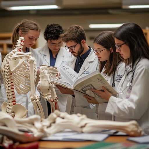
Why Dental Professionals Need it ? -- Dental Anatomy Models
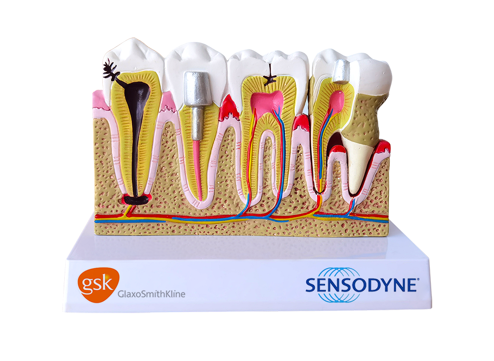
Why Dental Professionals Need it ? -- Dental Anatomy Models
Not just a display, but also an "immersive" learning experience about the Dental structure
Ushering in the "High-Definition Era" of Dental Anatomy
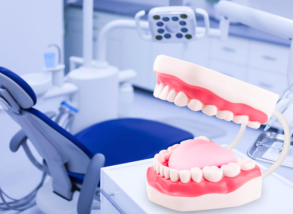
Part 1. Ingenious Anatomical Details
High-magnification and Authenticity: the tooth models are typically several times larger than real teeth( e.g.3x or 5x), allowing for a clear view of the teeth structures. Enlarged tooth model is really great dental teaching model.
Detailed description: Detailed depiction of the enamel, dentin, pulp cavity, root canal, and periodontal tissues , etc.., even able to show the shapes of common dental conditions (e.g., caries,periodontitis, periapical abscesses, fillings,and crowns). These anatomical tooth models are more suitable for dental students to learn.
Removable Feature: the anatomical tooth model is detachable and reassembled, completely used for experiments and exercises. Removable tooth teaching tools clearly show the lesions of caries,wisdom teeth,pits and fissures,root canals and root apex,etc., enabling teaching and making the learning more interactive and engaging. The root canal section of the Dental Implant Study Analysis demonstration teeth model is clearly displayed, which is convenient for doctor-patient communication.
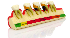
Part 2. Multi-scenario Applications
Dental Education and Research
For Dental college students, newly trained dental practitioners.
Clinical Doctor-Patient Communication
For dental clinicians / hospitals
Public Education and Health Education
For Dentists at dental clinics / community / school / oral health awareness campaign.
How to Perform a Breast Self-Exam (BSE) — Using Breast Models to Support Early Detection
The Importance of Anatomical Models and Medical Training Simulators in Medical Education